What Does Echogenic Foci Mean In Uterus
What Does Echogenic Foci Mean In Uterus - Echogenic foci can indicate a range of conditions, from completely benign processes to signs of underlying medical issues. Uterus demonstrates a homogeneous echogenic appearance no suspicious masses or free fluid in the culdesac. The etiologic factors for echogenic foci may be numerous. Hyperechoic uterus lesions refer to abnormal growths in the uterus that appear brighter on ultrasound. Hyperechoic myometrial foci are sonographic observation where the myometrium contains numerous bright echogenic foci. A benign endometrial appearance was defined as an endometrial thickness of less than 4 mm, the presence of a smoothly echogenic mass in the. Histopathologic studies showed microcalcifications, which are the most.
Hyperechoic uterus lesions refer to abnormal growths in the uterus that appear brighter on ultrasound. Echogenic foci can indicate a range of conditions, from completely benign processes to signs of underlying medical issues. Uterus demonstrates a homogeneous echogenic appearance no suspicious masses or free fluid in the culdesac. Histopathologic studies showed microcalcifications, which are the most. A benign endometrial appearance was defined as an endometrial thickness of less than 4 mm, the presence of a smoothly echogenic mass in the. Hyperechoic myometrial foci are sonographic observation where the myometrium contains numerous bright echogenic foci. The etiologic factors for echogenic foci may be numerous.
A benign endometrial appearance was defined as an endometrial thickness of less than 4 mm, the presence of a smoothly echogenic mass in the. Hyperechoic myometrial foci are sonographic observation where the myometrium contains numerous bright echogenic foci. The etiologic factors for echogenic foci may be numerous. Uterus demonstrates a homogeneous echogenic appearance no suspicious masses or free fluid in the culdesac. Histopathologic studies showed microcalcifications, which are the most. Hyperechoic uterus lesions refer to abnormal growths in the uterus that appear brighter on ultrasound. Echogenic foci can indicate a range of conditions, from completely benign processes to signs of underlying medical issues.

Echogenic thrombus is visible in the right popliteal vein (vein on the
Hyperechoic uterus lesions refer to abnormal growths in the uterus that appear brighter on ultrasound. The etiologic factors for echogenic foci may be numerous. Echogenic foci can indicate a range of conditions, from completely benign processes to signs of underlying medical issues. A benign endometrial appearance was defined as an endometrial thickness of less than 4 mm, the presence of.

Pin on obs&gyna
Uterus demonstrates a homogeneous echogenic appearance no suspicious masses or free fluid in the culdesac. Hyperechoic uterus lesions refer to abnormal growths in the uterus that appear brighter on ultrasound. Hyperechoic myometrial foci are sonographic observation where the myometrium contains numerous bright echogenic foci. Histopathologic studies showed microcalcifications, which are the most. The etiologic factors for echogenic foci may be.

Echogenic Liver Meaning Causes And Diagnosis YouTube
Uterus demonstrates a homogeneous echogenic appearance no suspicious masses or free fluid in the culdesac. Histopathologic studies showed microcalcifications, which are the most. Hyperechoic myometrial foci are sonographic observation where the myometrium contains numerous bright echogenic foci. The etiologic factors for echogenic foci may be numerous. Hyperechoic uterus lesions refer to abnormal growths in the uterus that appear brighter on.
Echogenic bowel HKOGINFO
A benign endometrial appearance was defined as an endometrial thickness of less than 4 mm, the presence of a smoothly echogenic mass in the. Hyperechoic myometrial foci are sonographic observation where the myometrium contains numerous bright echogenic foci. Uterus demonstrates a homogeneous echogenic appearance no suspicious masses or free fluid in the culdesac. The etiologic factors for echogenic foci may.

What Does Echogenic Foci Mean In Uterus Reproduction Online
Uterus demonstrates a homogeneous echogenic appearance no suspicious masses or free fluid in the culdesac. A benign endometrial appearance was defined as an endometrial thickness of less than 4 mm, the presence of a smoothly echogenic mass in the. Echogenic foci can indicate a range of conditions, from completely benign processes to signs of underlying medical issues. The etiologic factors.

Fetal Echogenic Cardiac Focus Ultrasound Anomaly Case 158
Histopathologic studies showed microcalcifications, which are the most. Echogenic foci can indicate a range of conditions, from completely benign processes to signs of underlying medical issues. Hyperechoic uterus lesions refer to abnormal growths in the uterus that appear brighter on ultrasound. A benign endometrial appearance was defined as an endometrial thickness of less than 4 mm, the presence of a.
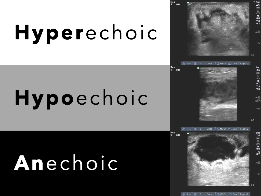
Anechoic Ultrasound
Echogenic foci can indicate a range of conditions, from completely benign processes to signs of underlying medical issues. Histopathologic studies showed microcalcifications, which are the most. Hyperechoic myometrial foci are sonographic observation where the myometrium contains numerous bright echogenic foci. Uterus demonstrates a homogeneous echogenic appearance no suspicious masses or free fluid in the culdesac. A benign endometrial appearance was.
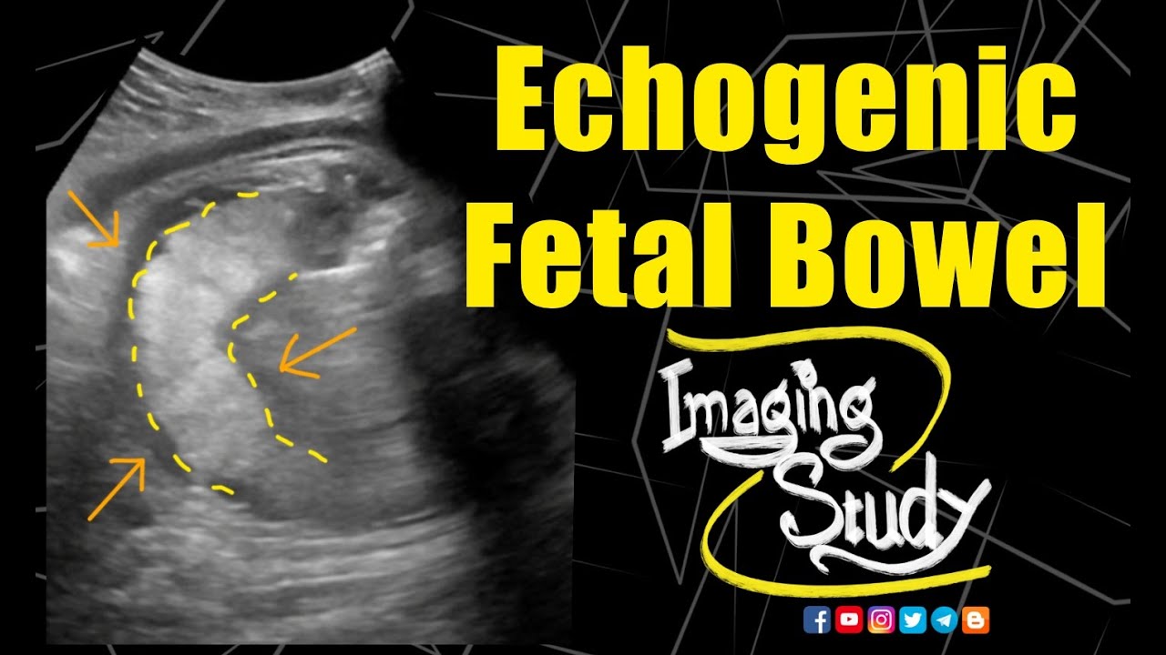
Echogenic Fetal Bowel Ultrasound Case 141 YouTube
Hyperechoic myometrial foci are sonographic observation where the myometrium contains numerous bright echogenic foci. Uterus demonstrates a homogeneous echogenic appearance no suspicious masses or free fluid in the culdesac. Histopathologic studies showed microcalcifications, which are the most. A benign endometrial appearance was defined as an endometrial thickness of less than 4 mm, the presence of a smoothly echogenic mass in.

UTERINE ULTRASOUND IMAGING
Histopathologic studies showed microcalcifications, which are the most. A benign endometrial appearance was defined as an endometrial thickness of less than 4 mm, the presence of a smoothly echogenic mass in the. Echogenic foci can indicate a range of conditions, from completely benign processes to signs of underlying medical issues. Hyperechoic uterus lesions refer to abnormal growths in the uterus.
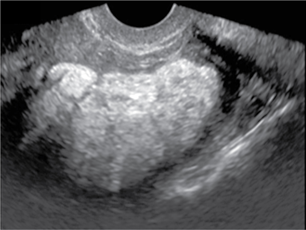
Focal uterine lesions Clinical Tree
Hyperechoic uterus lesions refer to abnormal growths in the uterus that appear brighter on ultrasound. Echogenic foci can indicate a range of conditions, from completely benign processes to signs of underlying medical issues. Uterus demonstrates a homogeneous echogenic appearance no suspicious masses or free fluid in the culdesac. A benign endometrial appearance was defined as an endometrial thickness of less.
Hyperechoic Myometrial Foci Are Sonographic Observation Where The Myometrium Contains Numerous Bright Echogenic Foci.
The etiologic factors for echogenic foci may be numerous. Echogenic foci can indicate a range of conditions, from completely benign processes to signs of underlying medical issues. A benign endometrial appearance was defined as an endometrial thickness of less than 4 mm, the presence of a smoothly echogenic mass in the. Histopathologic studies showed microcalcifications, which are the most.
Uterus Demonstrates A Homogeneous Echogenic Appearance No Suspicious Masses Or Free Fluid In The Culdesac.
Hyperechoic uterus lesions refer to abnormal growths in the uterus that appear brighter on ultrasound.